Glaucoma Testing for the Most Accurate Results: Standard vs. Supplemental
Blog:Glaucoma Testing for the Most Accurate Results: Standard vs. Supplemental
Glaucoma Testing for the Most Accurate Results: Standard vs. Supplemental
Glaucoma Testing for the Most Accurate Results: Standard vs. Supplemental
Glaucoma is a serious eye condition that can lead to permanent vision loss if left undiagnosed or untreated. Because glaucoma often progresses without noticeable symptoms in its early stages, regular and precise testing is critical for early detection. At Texas State Optical, we utilize a combination of standard and supplemental tests to ensure the most accurate diagnosis and monitoring of glaucoma. Understanding the differences between these tests can help patients make informed decisions about their eye health.
The Importance of Regular Eye Exams
Regular eye exams are essential for maintaining eye health and detecting serious conditions like glaucoma in their early stages. Many eye diseases, including glaucoma, develop gradually and may not present noticeable symptoms until significant vision loss has occurred. Through comprehensive eye exams, optometrists can identify subtle changes in eye pressure, optic nerve health, and visual field function before irreversible damage takes place.
Early detection allows for timely intervention, which can slow or prevent vision loss with treatments such as medication, laser therapy, or surgery. Annual eye exams not only help preserve sight but also contribute to overall well-being by detecting other health conditions, such as diabetes and high blood pressure, which often manifest in the eyes.
Standard Glaucoma Tests
Standard glaucoma tests are commonly performed during routine eye exams to assess the risk of glaucoma and detect early signs of the disease. These include:
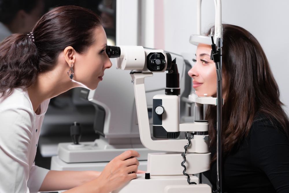
• Tonometry (Eye Pressure Test): Tonometry measures intraocular pressure (IOP), which is one of the primary risk factors for glaucoma. The most common method is the Goldmann applanation tonometry, which uses a small probe to gently flatten the cornea and measure pressure. A non-contact version, the air puff test, is also used but may be less precise. Elevated IOP can indicate glaucoma, but pressure alone is not enough to confirm a diagnosis.
• Ophthalmoscopy (Optic Nerve Examination): An eye doctor examines the optic nerve for signs of damage using a specialized magnifying lens. This test allows for a visual assessment of the nerve’s shape, color, and cupping (hollowing at the center), which may indicate glaucoma-related damage.
Supplemental Glaucoma Tests for Greater Accuracy
In addition to standard tests, supplemental testing provides a more detailed assessment, especially for patients at higher risk or those with inconclusive results. These include:
• Visual Field Test (Perimetry): This test maps the patient’s field of vision to identify any blind spots or vision loss that may be associated with glaucoma. It helps determine how much peripheral vision has been affected, as glaucoma typically damages side vision before central vision.
• Pachymetry (Corneal Thickness Measurement): Since corneal thickness can influence eye pressure readings, a pachymetry test measures the thickness of the cornea to help interpret IOP readings accurately. Thinner corneas may lead to underestimated pressure readings, while thicker corneas may cause overestimated results.
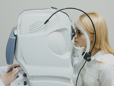
• Optical Coherence Tomography (OCT): OCT is an advanced imaging test that provides high-resolution cross-sectional images of the retina and optic nerve. It measures the thickness of the retinal nerve fiber layer, allowing for early detection of glaucoma-related thinning before vision loss occurs.
• Gonioscopy (Angle Examination): Gonioscopy evaluates the drainage angle of the eye to determine whether a patient has open-angle or angle-closure glaucoma. This test is essential for understanding how fluid exits the eye and identifying abnormalities that could contribute to elevated IOP.
• Fundus Photography (Optic Nerve Imaging): High-resolution digital images of the optic nerve allow for detailed comparisons over time. These images help track subtle changes in the optic nerve that might indicate glaucoma progression.
Schedule Your Glaucoma Screening Today
Glaucoma is a silent but serious threat to vision. Relying on a combination of standard and supplemental tests ensures early detection and more precise monitoring of the condition. At Texas State Optical, we use advanced diagnostic tools to assess your eye health and create personalized treatment plans.
If you are at risk for glaucoma, contact Texas State Optical to schedule an eye exam and protect your vision for the future. Visit our office in Texas City, Texas, or call (409) 202-6984 to book an appointment today.
Quick Links
Helpful Articles
article_category: cosmetic
article_category: eye surgery co-management
article_category: products
article_category: vision therapy
article_category: dogs
article_category: technology
article_category: services
article_category: general
article_category: FAQ & tips
article_category: eye health
article_category: aesthetics
article_category: exotic
article_category: large animal
article_category: contact lenses
article_category: surgical procedures
article_category: health
article_category: psychiatry
article_category: conditions
article_category: restorative
article_category: preventative
article_category: cats
article_category: eyeglasses
article_category: ocular disease management
[]
Flowbite is an open-source library of interactive components built on top of Tailwind CSS including buttons, dropdowns, modals, navbars, and more.
Check out this guide to learn how to get started and start developing websites even faster with components on top of Tailwind CSS.
All Eyecare Services
We offer a wide variety of eye care services to the Texas City community. Contact us with any questions about our services.
Keep In Touch
For non-urgent questions or to learn more about our services, contact us today!
Contact Information
Office Hours
- Monday 8:00am - 5:30pm
- Tuesday 8:00am - 5:30pm
- Wednesday 8:00am - 5:30pm
- Thursday 8:00am - 5:30pm
- Friday 8:00am - 5:30pm
- Saturday 8:00am - 1:00pm
- Sunday Closed
- Monday
- Tuesday
- Wednesday
- Thursday
- Friday
- Saturday
- Sunday
- Monday
- Tuesday
- Wednesday
- Thursday
- Friday
- Saturday
- Sunday
© 2025 Texas State Optical. All rights Reserved - Accessibility Statement - Privacy Policy - Sitemap
Managed and Designed by
![]()